 English
 العربية
 Deutsch
 Español
 Français
 हिन्दी
 Bahasa Indonesia
 Italiano
 日本語
日本語 한국어
한국어 Polski
 Português (Brasil)
 Русский
 Türkçe
 Українська
 简体中文
简体中文Learn
How small is a cell?

A human liver cell averages 25 µm across, while the giant amoeba Chaos carolinense can stretch to 1 mm long. There is no single cell size: familiar human cells can be tens of micrometres across, while some single-celled organisms grow large enough to appear as moving specks.
A micrometre (µm) is a length unit used for cells; a nanometre (nm) is smaller. Those units make it easier to follow the shift from a human cell to a bacterium, then to a tiny structure inside a cell.
A human cell can be close to the width of a hair
A human hair is about 70 µm wide. A cheek cell measures 60 µm across, nearly hair-width, and about eight red blood cell diameters across. A red blood cell is 7.5 µm across. These familiar human cell types put “microscopic” into perspective: the cheek cell is close to the width of a hair, while a red blood cell is far narrower.
Cheek cells are flat, squamous cells from the lining inside the mouth. Their broad shape lets them form a surface lining, and they are easy to collect with a cheek swab. That makes them a common sample for classroom DNA extraction experiments. The sample starts with a part of the body most people can reach, but the cells themselves need a microscope to see clearly.
A mature human adipocyte, or fat cell, has a diameter of 100 µm. Almost its entire interior is filled by a single large lipid droplet, which pushes the nucleus to the edge. At 100 µm, it is about 1.7 times as wide as a cheek cell. The large droplet gives this cell a distinctive layout: a stored reserve fills most of the space inside its outer membrane.
A hepatocyte, a liver cell, averages 25 µm across. It is smaller than a cheek cell but about 3.3 red blood cell diameters across. Hepatocytes make up about 80% of the liver’s mass and perform hundreds of metabolic functions. A cell’s width alone does not tell you what work it does: the smaller hepatocyte is part of an organ responsible for many kinds of chemical activity.
Some single-celled organisms are much larger
Chaos carolinense is one of the largest known amoebae and can look like a moving speck to the naked eye. When extended, it measures 1 mm long, about twice the width of a medium grain of sand. A grain of rice is about six times as long as the extended amoeba. The amoeba’s outline shifts as it moves, so its measured length describes its extended form.
The pink ciliate Blepharisma japonicum measures 300 µm long, about 4.3 human-hair widths. That is also about 40 red blood cell diameters. Blepharisma’s light-sensitive pigment, blepharismin, gives it its pink color and can be toxic in bright light. A cell this large is still too small to see clearly without magnification, but it is many times wider than the blood cell that helps carry oxygen around your body.
Bacteria shrink the scale to a few micrometres
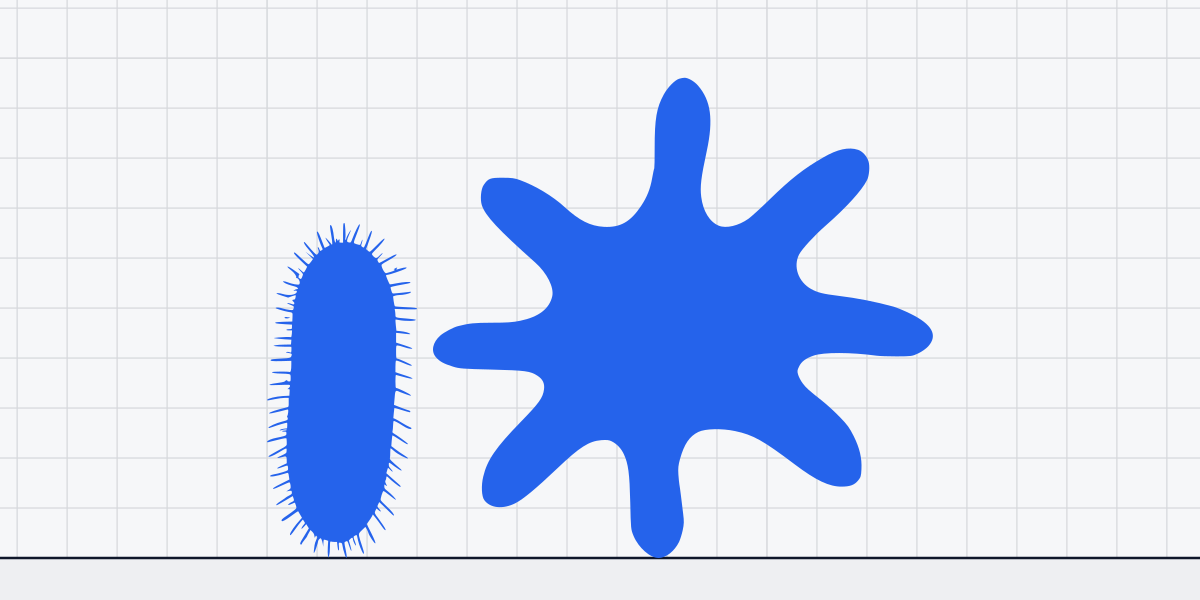
Bacteria are cells, but they are much smaller than the human cells above. The rod-shaped bacterium Shigella flexneri measures 2.5 µm long. A red blood cell spans about three Shigella lengths. Shigella can invade human intestinal cells using a specialized injection system. Its small dimensions do not prevent it from interacting directly with much larger cells.
Deinococcus radiodurans measures 2 µm across. It can survive radiation doses thousands of times higher than would kill a human by rapidly repairing shattered DNA. A red blood cell is about 3.8 times its diameter. This bacterium takes up only a small space beside a blood cell, yet its repair system can restore damaged genetic material.
The marine cyanobacterium Synechococcus averages 1 µm across. A red blood cell is about 7.5 times wider, and Synechococcus is about half the diameter of Deinococcus radiodurans. Synechococcus is one of the most abundant photosynthetic organisms on Earth and contributes significantly to global oxygen production. Its role reaches far beyond its tiny outline: these cells help drive photosynthesis in the oceans.
Viruses are smaller than cells, but are not cells
Among the smallest known cells, Mycoplasma genitalium measures about 0.3 by 0.6 micrometres (opens in a new tab). Smaller structures also exist inside cells. A lysosome is a membrane-bound organelle containing digestive enzymes that break down waste and cellular debris; its typical diameter is 500 nm. That is about half the diameter of Synechococcus. A lysosome is part of a cell, not an independent cell of its own.
A Lassa virus particle measures 100 nm across, about one-fifth the diameter of a lysosome. Viruses cannot reproduce on their own; they use a host cell’s machinery. The distinction matters even at this scale: a virus may be smaller than a cell, but it does not carry out reproduction independently.
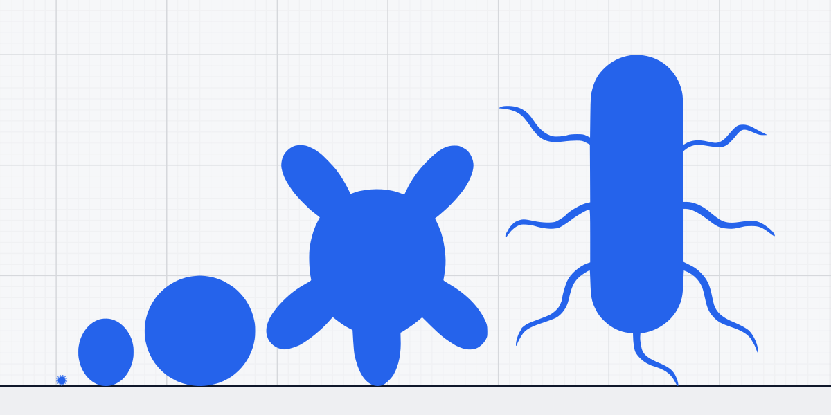
A red blood cell spans about 75 Lassa virus diameters, yet the virus still needs a host cell to reproduce.
Sources
- McGowin & Totten, "The Unique Microbiology and Molecular Pathogenesis of Mycoplasma genitalium," J Infect Dis 2017 (PMC5853509) (opens in a new tab) pmc.ncbi.nlm.nih.gov
- ViralZone, SIB Swiss Institute of Bioinformatics; CDC Poxvirus overview
- CDC: Gonorrhea; Kenneth Todar's Microbiology
- CDC: Listeria; ASM Microbe Library
- CDC Fungal Diseases; Mycology textbooks
- Encyclopedia Britannica: Chlorella; Algae Research textbook
- Alberts, Molecular Biology of the Cell; Kuby Immunology
- ASM: Fungal spore morphology; Mycology textbooks
- Kumar, Robbins Basic Pathology; Alberts, Molecular Biology of the Cell
- Molecular Biology of the Cell, Alberts et al.; Histology Guide
- Physiological Reviews: Adipocyte biology; Molecular Biology of the Cell
- Protist Information Server, Didinium
- Protist Information Server - Chaos carolinense
- Protist Information Server: Blepharisma; Encyclopedia of Life: Blepharisma japonicum
- Histology Guide: buccal epithelium
- Alberts, Molecular Biology of the Cell; Histology Atlas
- CDC: Shigella; ASM Microbe Library
- ASM Journals: Deinococcus radiodurans review; NCBI: Deinococcus genome
- NCBI Genome; Nature Reviews Microbiology - Marine Cyanobacteria
- Molecular Biology of the Cell, 6th ed.